xtal

At the GLUPOR12

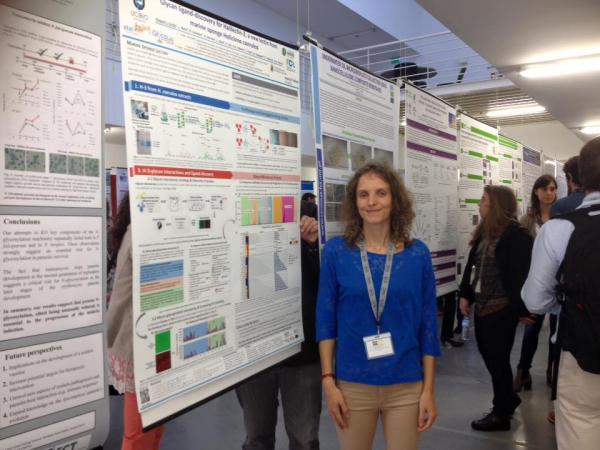
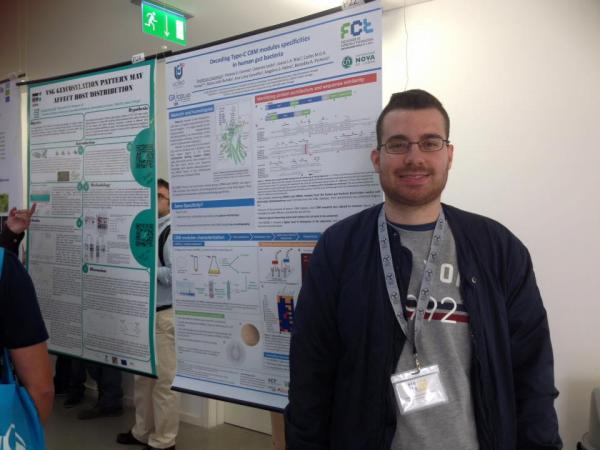
Raquel Costa and Frederico Lourenço are presenting their research work as poster communications at Glupor12, in Aveiro!

Polino, M, Carvalho AL, Juknaitė L, Portugal CAM, Coelhoso IM, Romão MJ, Crespo JG.  2017.  Ion-Exchange Membranes for Stable Derivatization of Protein Crystals, 2017. Crystal Growth & DesignCrystal Growth & Design. : American Chemical Society AbstractWebsite
n/a

New paper online!

Ion-Exchange Membranes for Stable Derivatization of Protein Crystals

Otrelo-Cardoso, AR, Nair RR, Correia MAS, Cordeiro RCS, Panjkovich A, Svergun DI, Santos-Silva T, Rivas MG.  2017.  Highly selective tungstate transporter protein TupA from Desulfovibrio alaskensis G20, 2017. Scientific Reports. 7(1):5798. AbstractWebsite

Molybdenum and tungsten are taken up by bacteria and archaea as their soluble oxyanions through high affinity transport systems belonging to the ATP-binding cassette (ABC) transporters. The component A (ModA/TupA) of these transporters is the first selection gate from which the cell differentiates between MoO4 2−, WO4 2− and other similar oxyanions. We report the biochemical characterization and the crystal structure of the apo-TupA from Desulfovibrio desulfuricans G20, at 1.4 Å resolution. Small Angle X-ray Scattering data suggests that the protein adopts a closed and more stable conformation upon ion binding. The role of the arginine 118 in the selectivity of the oxyanion was also investigated and three mutants were constructed: R118K, R118E and R118Q. Isothermal titration calorimetry clearly shows the relevance of this residue for metal discrimination and oxyanion binding. In this sense, the three variants lost the ability to coordinate molybdate and the R118K mutant keeps an extremely high affinity for tungstate. These results contribute to an understanding of the metal-protein interaction, making it a suitable candidate for a recognition element of a biosensor for tungsten detection.

New paper online!

Highly selective tungstate transporter protein TupA from Desulfovibrio alaskensis G20
Ana Rita Otrelo-Cardoso, Rashmi R. Nair, Márcia A. S. Correia, Raquel S. Correia Cordeiro, Alejandro Panjkovich, Dmitri I. Svergun, Teresa Santos-Silva & Maria G. Rivas 

Maria João Romão elected member of the Faculty's Council

Congratulations to Maria João and the other new members of the Faculty's Council, elected yesterday!

If you want to know more about the FCT-NOVA Council's duties: http://www.cf.fct.unl.pt

Romão, MJ, Coelho C, Santos-Silva T, Foti A, Terao M, Garattini E, Leimkühler S.  2017.  Structural basis for the role of mammalian aldehyde oxidases in the metabolism of drugs and xenobiotics. Current Opinion in Chemical Biology. 37:39-47. AbstractWebsite

Aldehyde oxidases (AOXs) are molybdo-flavoenzymes characterized by broad substrate specificity, oxidizing aromatic/aliphatic aldehydes into the corresponding carboxylic acids and hydroxylating various heteroaromatic rings. Mammals are characterized by a complement of species-specific \{AOX\} isoenzymes, that varies from one in humans (AOX1) to four in rodents (AOX1, AOX2, \{AOX3\} and AOX4). The physiological function of mammalian \{AOX\} isoenzymes is unknown, although human \{AOX1\} is an emerging enzyme in phase-I drug metabolism. Indeed, the number of therapeutic molecules under development which act as \{AOX\} substrates is increasing. The recent crystallization and structure determination of human \{AOX1\} as well as mouse \{AOX3\} has brought new insights into the mechanisms underlying substrate/inhibitor binding as well as the catalytic activity of this class of enzymes.

Kowacz, M, Marchel M, Juknaité L, Esperança JMSS, Romão MJ, Carvalho AL, Rebelo LPN.  2017.  Infrared light-induced protein crystallization. Structuring of protein interfacial water and periodic self-assembly. Journal of Crystal Growth. 457:362-368. AbstractWebsite

Abstract We show that a physical trigger, a non-ionizing infrared (IR) radiation at wavelengths strongly absorbed by liquid water, can be used to induce and kinetically control protein (periodic) self-assembly in solution. This phenomenon is explained by considering the effect of İR\} light on the structuring of protein interfacial water. Our results indicate that the İR\} radiation can promote enhanced mutual correlations of water molecules in the protein hydration shell. We report on the radiation-induced increase in both the strength and cooperativeness of H-bonds. The presence of a structured dipolar hydration layer can lead to attractive interactions between like-charged biomacromolecules in solution (and crystal nucleation events). Furthermore, our study suggests that enveloping the protein within a layer of structured solvent (an effect enhanced by İR\} light) can prevent the protein non-specific aggregation favoring periodic self-assembly. Recognizing the ability to affect protein-water interactions by means of İR\} radiation may have important implications for biological and bio-inspired systems.

Pires, VMR, Pereira PMM, Brás JLA, Correia M, Cardoso V, Bule P, Alves VD, Najmudin S, Venditto I, Ferreira LMA, Romão MJ, Carvalho AL, Fontes CMGA, Prazeres DM.  2017.  Stability and ligand promiscuity of type A carbohydrate-binding modules are illustrated by the structure of Spirochaeta thermophila StCBM64C, mar. Journal of Biological Chemistry. 292:4847–4860., Number 12 AbstractWebsite

Deconstruction of cellulose, the most abundant plant cell wall polysaccharide, requires the cooperative activity of a large repertoire of microbial enzymes. Modular cellulases contain non-catalytic type A Carbohydrate-Binding Modules (CBMs) that specifically bind to the crystalline regions of cellulose, thus promoting enzyme efficacy through proximity and targeting effects. Although type A CBMs play a critical role in cellulose recycling, their mechanism of action remains poorly understood. Here we produced a library of recombinant CBMs representative of the known diversity of type A modules. The binding properties of 40 CBMs, in fusion with an N-terminal green fluorescence protein (GFP) domain, revealed that type A CBMs possess the ability to recognize different crystalline forms of cellulose and chitin over a wide range of temperatures, pHs and ionic strengths. A Spirochaeta thermophila CBM64, in particular, displayed plasticity in its capacity to bind both crystalline and soluble carbohydrates under a wide range of extreme conditions. The structure of S. thermophila StCBM64C revealed an untwisted, flat, carbohydrate-binding interface comprising the side chains of four tryptophan residues in a coplanar linear arrangement. Significantly, two highly conserved asparagine side chains, each one located between two tryptophan residues, are critical to insoluble and soluble glucan recognition but not to bind xyloglucan. Thus, CBM64 compact structure and its extended and versatile ligand interacting platform illustrates how type A CBMs target their appended plant cell wall degrading enzymes to a diversity of recalcitrant carbohydrates under a wide range of environmental conditions.

Bule, P, Alves VD, Israeli-Ruimy V, Carvalho AL, Ferreira LMA, Smith SP, Gilbert HJ, Najmudin S, Bayer EA, Fontes CMGA.  2017.  Assembly of Ruminococcus flavefaciens cellulosome revealed by structures of two cohesin-dockerin complexes, 2017. Scientific Reports. 7:759. AbstractWebsite

Cellulosomes are sophisticated multi-enzymatic nanomachines produced by anaerobes to effectively deconstruct plant structural carbohydrates. Cellulosome assembly involves the binding of enzyme-borne dockerins (Doc) to repeated cohesin (Coh) modules located in a non-catalytic scaffoldin. Docs appended to cellulosomal enzymes generally present two similar Coh-binding interfaces supporting a dual-binding mode, which may confer increased positional adjustment of the different complex components. Ruminococcus flavefaciens’ cellulosome is assembled from a repertoire of 223 Doc-containing proteins classified into 6 groups. Recent studies revealed that Docs of groups 3 and 6 are recruited to the cellulosome via a single-binding mode mechanism with an adaptor scaffoldin. To investigate the extent to which the single-binding mode contributes to the assembly of R. flavefaciens cellulosome, the structures of two group 1 Docs bound to Cohs of primary (ScaA) and adaptor (ScaB) scaffoldins were solved. The data revealed that group 1 Docs display a conserved mechanism of Coh recognition involving a single-binding mode. Therefore, in contrast to all cellulosomes described to date, the assembly of R. flavefaciens cellulosome involves single but not dual-binding mode Docs. Thus, this work reveals a novel mechanism of cellulosome assembly and challenges the ubiquitous implication of the dual-binding mode in the acquisition of cellulosome flexibility.

Otrelo-Cardoso, AR, Nair RR, Correia MAS, Rivas MG, Santos-Silva T.  2014.  TupA: A Tungstate Binding Protein in the Periplasm of Desulfovibrio alaskensis G20, 2014/05/29/accep. International Journal of Molecular Sciences. 15(7):11783-11798.: MDPI AbstractWebsite

The TupABC system is involved in the cellular uptake of tungsten and belongs to the ABC (ATP binding cassette)-type transporter systems. The TupA component is a periplasmic protein that binds tungstate anions, which are then transported through the membrane by the TupB component using ATP hydrolysis as the energy source (the reaction catalyzed by the ModC component). We report the heterologous expression, purification, determination of affinity binding constants and crystallization of the Desulfovibrio alaskensis G20 TupA. The tupA gene (locus tag Dde_0234) was cloned in the pET46 Enterokinase/Ligation-Independent Cloning (LIC) expression vector, and the construct was used to transform BL21 (DE3) cells. TupA expression and purification were optimized to a final yield of 10 mg of soluble pure protein per liter of culture medium. Native polyacrylamide gel electrophoresis was carried out showing that TupA binds both tungstate and molybdate ions and has no significant interaction with sulfate, phosphate or perchlorate. Quantitative analysis of metal binding by isothermal titration calorimetry was in agreement with these results, but in addition, shows that TupA has higher affinity to tungstate than molybdate. The protein crystallizes in the presence of 30% (w/v) polyethylene glycol 3350 using the hanging-drop vapor diffusion method. The crystals diffract X-rays beyond 1.4 Å resolution and belong to the P2(1) space group, with cell parameters a = 52.25 Å, b = 42.50 Å, c = 54.71 Å, β = 95.43°. A molecular replacement solution was found, and the structure is currently under refinement.

dos Santos, R, Carvalho AL, Roque CAA.  2017.  Renaissance of protein crystallization and precipitation in biopharmaceuticals purification, 2017/1//. Biotechnology Advances. 35(1):41-50. AbstractWebsite

AbstractThe current chromatographic approaches used in protein purification are not keeping pace with the increasing biopharmaceutical market demand. With the upstream improvements, the bottleneck shifted towards the downstream process. New approaches rely in Anything But Chromatography methodologies and revisiting former techniques with a bioprocess perspective. Protein crystallization and precipitation methods are already implemented in the downstream process of diverse therapeutic biological macromolecules, overcoming the current chromatographic bottlenecks. Promising work is being developed in order to implement crystallization and precipitation in the purification pipeline of high value therapeutic molecules. This review focuses in the role of these two methodologies in current industrial purification processes, and highlights their potential implementation in the purification pipeline of high value therapeutic molecules, overcoming chromatographic holdups.

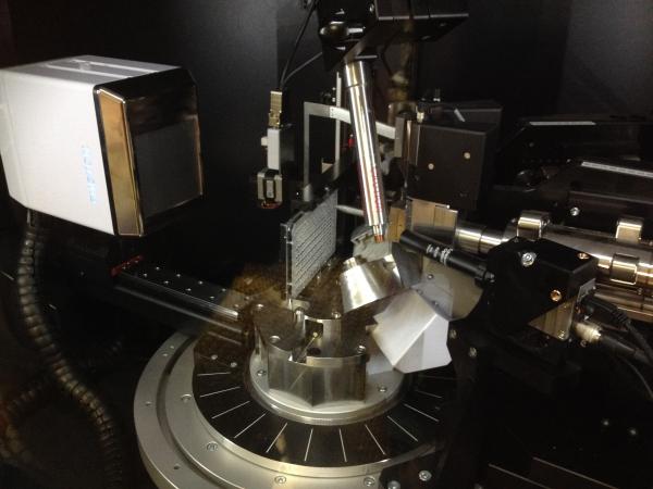
In situ diffraction is now possible in our X-ray diffractometer!

We are now equipped with a fully motorized ISX stage, which allows the fully automated screening of samples in crystallization plates.

Correia, MAS, Otrelo-Cardoso AR, Schwuchow V, {Sigfridsson Clauss} KGV, Haumann M, Romão MJ, Leimkühler S, Santos-Silva T.  2016.  {The Escherichia coli Periplasmic Aldehyde Oxidoreductase Is an Exceptional Member of the Xanthine Oxidase Family of Molybdoenzymes}, oct. ACS Chemical Biology. 11:2923–2935., Number 10 AbstractWebsite

The xanthine oxidase (XO) family comprises molybdenum-dependent enzymes that usually form homodimers (or dimers of heterodimers/trimers) organized in three domains that harbor two [2Fe-2S] clusters, one FAD, and a Mo cofactor. In this work, we crystallized an unusual member of the family, the periplasmic aldehyde oxidoreductase PaoABC from Escherichia coli. This is the first example of an E. coli protein containing a molybdopterin-cytosine-dinucleotide cofactor and is the only heterotrimer of the XO family so far structurally characterized. The crystal structure revealed the presence of an unexpected [4Fe-4S] cluster, anchored to an additional 40 residues subdomain. According to phylogenetic analysis, proteins containing this cluster are widely spread in many bacteria phyla, putatively through repeated gene transfer events. The active site of PaoABC is highly exposed to the surface with no aromatic residues and an arginine (PaoC-R440) making a direct interaction with PaoC-E692, which acts as a base catalyst. In order to understand the importance of R440, kinetic assays were carried out, and the crystal structure of the PaoC-R440H variant was also determined.

Brás, JLA, Pinheiro BA, Cameron K, Cuskin F, Viegas A, Najmudin S, Bule P, Pires VMR, Romão MJ, Bayer EA, Spencer HL, Smith S, Gilbert HJ, Alves VD, Carvalho AL, Fontes CMGA.  2016.  Diverse specificity of cellulosome attachment to the bacterial cell surface, dec. Scientific Reports. 6:38292.: The Author(s) AbstractWebsite

During the course of evolution, the cellulosome, one of Nature's most intricate multi-enzyme complexes, has been continuously fine-tuned to efficiently deconstruct recalcitrant carbohydrates. To facilitate the uptake of released sugars, anaerobic bacteria use highly ordered protein-protein interactions to recruit these nanomachines to the cell surface. Dockerin modules located within a non-catalytic macromolecular scaffold, whose primary role is to assemble cellulosomal enzymatic subunits, bind cohesin modules of cell envelope proteins, thereby anchoring the cellulosome onto the bacterial cell. Here we have elucidated the unique molecular mechanisms used by anaerobic bacteria for cellulosome cellular attachment. The structure and biochemical analysis of five cohesin-dockerin complexes revealed that cell surface dockerins contain two cohesin-binding interfaces, which can present different or identical specificities. In contrast to the current static model, we propose that dockerins utilize multivalent modes of cohesin recognition to recruit cellulosomes to the cell surface, a mechanism that maximises substrate access while facilitating complex assembly.

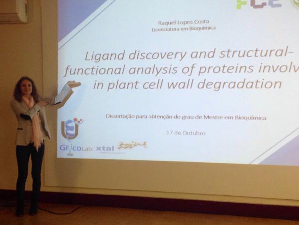
Congratulations, Raquel!

Raquel Costa successfully presented and defended her research work to obtain the MSc in Biochemistry.



Terao, M, Romão MJ, Leimkühler S, Bolis M, Fratelli M, Coelho C, Santos-Silva T, Garattini E.  2016.  Structure and function of mammalian aldehyde oxidases. Archives of Toxicology. 90:753–780., Number 4 AbstractWebsite

Mammalian aldehyde oxidases (AOXs; EC1.2.3.1) are a group of conserved proteins belonging to the family of molybdo-flavoenzymes along with the structurally related xanthine dehydrogenase enzyme. AOXs are characterized by broad substrate specificity, oxidizing not only aromatic and aliphatic aldehydes into the corresponding carboxylic acids, but also hydroxylating a series of heteroaromatic rings. The number of AOX isoenzymes expressed in different vertebrate species is variable. The two extremes are represented by humans, which express a single enzyme (AOX1) in many organs and mice or rats which are characterized by tissue-specific expression of four isoforms (AOX1, AOX2, AOX3, and AOX4). In vertebrates each AOX isoenzyme is the product of a distinct gene consisting of 35 highly conserved exons. The extant species-specific complement of AOX isoenzymes is the result of a complex evolutionary process consisting of a first phase characterized by a series of asynchronous gene duplications and a second phase where the pseudogenization and gene deletion events prevail. In the last few years remarkable advances in the elucidation of the structural characteristics and the catalytic mechanisms of mammalian AOXs have been made thanks to the successful crystallization of human AOX1 and mouse AOX3. Much less is known about the physiological function and physiological substrates of human AOX1 and other mammalian AOX isoenzymes, although the importance of these proteins in xenobiotic metabolism is fairly well established and their relevance in drug development is increasing. This review article provides an overview and a discussion of the current knowledge on mammalian AOX.

Foti, A, Hartmann T, Coelho C, Santos-Silva T, Romão MJ, Leimkühler S.  2016.  Optimization of the Expression of Human Aldehyde Oxidase for Investigations of Single-Nucleotide Polymorphisms. Drug Metabolism and Disposition. 44:1277–1285., Number 8: American Society for Pharmacology and Experimental Therapeutics AbstractWebsite

Aldehyde oxidase (AOX1) is an enzyme with broad substrate specificity, catalyzing the oxidation of a wide range of endogenous and exogenous aldehydes as well as N-heterocyclic aromatic compounds. In humans, the enzyme’s role in phase I drug metabolism has been established and its importance is now emerging. However, the true physiologic function of AOX1 in mammals is still unknown. Further, numerous single-nucleotide polymorphisms (SNPs) have been identified in human AOX1. SNPs are a major source of interindividual variability in the human population, and SNP-based amino acid exchanges in AOX1 reportedly modulate the catalytic function of the enzyme in either a positive or negative fashion. For the reliable analysis of the effect of amino acid exchanges in human proteins, the existence of reproducible expression systems for the production of active protein in ample amounts for kinetic, spectroscopic, and crystallographic studies is required. In our study we report an optimized expression system for hAOX1 in Escherichia coli using a codon-optimized construct. The codon-optimization resulted in an up to 15-fold increase of protein production and a simplified purification procedure. The optimized expression system was used to study three SNPs that result in amino acid changes C44W, G1269R, and S1271L. In addition, the crystal structure of the S1271L SNP was solved. We demonstrate that the recombinant enzyme can be used for future studies to exploit the role of AOX in drug metabolism, and for the identification and synthesis of new drugs targeting AOX when combined with crystallographic and modeling studies.

Congratulations, Doctor Marino Santos!

Yesterday, Marino Santos successfully presented and defended his PhD work, entitled "​Structural and Func