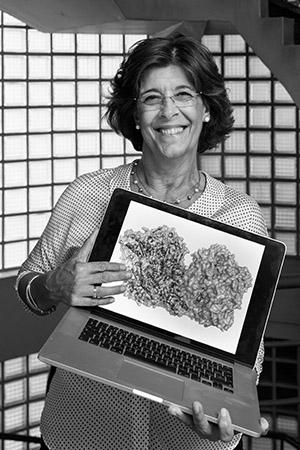
xtal

Coelho, C, Foti A, Hartmann T, Santos-Silva T, Leimk S, Rom MJ.  2015.  Structural insights into xenobiotic and inhibitor binding to human aldehyde oxidase, 2015. Nat Chem Biol. 11(10):779-83.Website

Structural Biology tools

(Inspired from Pereda's Lab)

Crystallizability predictors :)

Kowacz, M, Mukhopadhyay A, Carvalho AL, Esperanca J, Romao MJ, Rebelo LPN.  2012.  Hofmeister effects of ionic liquids in protein crystallization: Direct and water-mediated interactions. Crystengcomm. 14:4912-4921., Number 15 AbstractWebsite

We have performed experiments on the crystallization of two low molecular weight, positively charged proteins, lysozyme and ribonuclease A, using ionic liquids as either crystallization additives or, in particular cases, as precipitating agents. The ionic liquids (ILs) have been ordered according to their salting-in/out ability and the relative position of these ionic liquids in this ranking has been rationalized by considering their hydration properties (positive-negative, hydrophobic-hydrophilic). The ability to screen the effective charge of cationic proteins and aid protein nucleation (salting-out) has been shown to be superior for large polarizable anions with low charge density, negatively hydrated-Cl-, Br-, [SCN](-), methane-[C1SO3](-) and ethanesulfonates [C2SO3](-), than for anions with a relatively stable hydration shell, positively hydrated-lactate [Lac](-), butylsulfonate [C4SO3](-) and acetate [Ac](-). Upon increasing the background salt concentration, where electrostatic interactions are already effectively screened, the ability of the IL ions to stabilize proteins in solution (salting-in) has been shown to increase as the ions are likely to migrate to the non-polar protein surface and lower protein-water interfacial tension. This tendency is enhanced as the focus moves from those ions with positively hydrated hydrophilic compartments (e. g. [Ac](-)) to those with negatively hydrated groups (e. g. [C1SO3](-)) and the prevailing hydrophobic hydration (e. g. [C4SO3](-)). The observed inversion in the relative effect of ILs on protein crystallization with increasing ionic strength of the aqueous media has been interpreted as the differing effects of ion adsorption: charge screening and interfacial tension modification. Moreover, this work can further help in our understanding of the influence of ionic liquids on conformational changes of biomacromolecules in solution. Identification of the specific incorporation sites for choline and acetate ions, localized in two lysozyme crystals grown in pure IL solutions without any buffer or inorganic precipitant, can give us some insight into the role of the ionic liquid ions in protein structure development.

Ribeiro, D, Kulakova A, Quaresma P, Pereira E, Bonifacio C, Romao MJ, Franco R, Carvalho AL.  2014.  Use of Gold Nanoparticles as Additives in Protein Crystallization. Crystal Growth & Design. 14:222-227., Number 1 AbstractWebsite

Gold nanoparticles (AuNPs) exhibit unique properties that have made them a very attractive material for application in biological assays. Given the potentially interesting interactions between AuNPs and biological macromolecules, we investigated AuNPs-induced protein crystal growth. Differently functionalized AuNPs were tested as additives in cocrystallization studies with model proteins (hen egg white lysozyme (HEWL), ribonuclease A (RNase A), and proteinase K) as well as with case studies where there were problems in obtaining well-diffracting crystals. Trials were performed considering different crystallization drawbacks, from total absence of crystals to improvement of crystal morphology, size, twinning, and number of crystals per drop. Improvement of some of these factors was observed in the cases of HEWL, RNase A, phenylalanine hydroxylase (PAR), myoglobin, native aldehyde oxidase (AOH), and human albumin. In these proteins, the presence of the AuNPs promoted an increase in the size and/or better crystal morphology. From the systematic trials and subsequent observations, it can be concluded that the introduction of AuNPs should definitely be considered in crystal optimization trials to improve previously determined crystallization conditions.

Kowacz, M, Marchel M, Juknaite L, Esperanca J, Romao MJ, Carvalho AL, Rebelo LPN.  2015.  Ionic-Liquid-Functionalized Mineral Particles for Protein Crystallization. Crystal Growth & Design. 15:2994-3003., Number 6 AbstractWebsite

Nucleation is a critical step determining the outcome of the entire crystallization process. Finding an effective nucleant for protein crystallization is of utmost importance for structural biology. The latter relies on good-quality crystals to solve the three-dimensional structures of macromolecules. In this study we show that crystalline barium sulfate (BaSO4) with an etched and/or ionic liquid (IL)-functionalized surface (1) can induce protein nucleation at concentrations well below the concentration needed to promote crystal growth under control conditions, (2) can shorten the nucleation time, (3) can increase the growth rate, and finally (4) may help to improve the protein crystal morphology. These effects were shown for lysozyme, RNase A, trypsin, proteinase K, myoglobin, and hemoglobin. Therefore, the use of BaSO4 particles enables us to reduce the amount of protein in crystallization trials and increases the chance of obtaining protein crystals of the desired quality. In the context of the underlying mechanism, it is shown that the protein-solid contact formation is governed by the interaction of the polar compartments of the biomacromolecule with the support. The tendency of a protein to concentrate near the solid surface is enhanced by both the hydrophobicity of the protein and that of the surface (tuned by the functionalizing IL). These mechanisms of interaction of biomacromolecules with inorganic hydrophilic solids correspond to the principles of amphiphilic IL-mineral interactions.

Best Poster Award at GLUPOR11

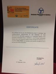
Diana RIbeiro was selected by the Scientific Committeee of the 11th International Meeting of the Portuguese Carbohydrate Group (GLUPOR11) & 6th Iberian Carbohydrate Meeting, as one of the three best poster presentations. The meeting was held at the Polytechnic Institute of Viseu, from the 6th to the 10th of September, 2015.

We hit the news!

Seixas, JD, Santos MFA, Mukhopadhyay A, Coelho AC, Reis PM, Veiros LF, Marques AR, Penacho N, Goncalves AML, Romao MJ, Bernardes GJL, Santos-Silva T, Romao CC.  2015.  A contribution to the rational design of Ru(CO)(3)Cl2L complexes for in vivo delivery of CO. Dalton Transactions. 44:5058-5075., Number 11 AbstractWebsite

A few ruthenium based metal carbonyl complexes, e.g. CORM-2 and CORM-3, have therapeutic activity attributed to their ability to deliver CO to biological targets. In this work, a series of related complexes with the formula [Ru(CO)(3)Cl2L] (L = DMSO (3), L-H3CSO(CH2)(2)CH(NH2)CO2H) (6a); D,L-H3CSO(CH2)(2)CH-(NH2)CO2H (6b); 3-NC5H4(CH2)(2)SO3.Na (7); 4-NC5H4(CH2)(2)SO3Na (8); PTA (9); DAPTA (10); H3CS-(CH2)(2)CH(OH) CO2H (11); CNCMe2CO2Me (12); CNCMeEtCO2Me (13); CN(c-C3H4)CO2Et) (14)) were designed, synthesized and studied. The effects of L on their stability, CO release profile, cytotoxicity and anti-inflammatory properties are described. The stability in aqueous solution depends on the nature of L as shown using HPLC and LC-MS studies. The isocyanide derivatives are the least stable complexes, and the S-bound methionine oxide derivative is the more stable one. The complexes do not release CO gas to the headspace, but release CO2 instead. X-ray diffraction of crystals of the model protein Hen Egg White Lysozyme soaked with 6b (4UWN) and 8 (4UWV) shows the addition of Ru-II(CO)(H2O)(4) at the His15 binding site. Soakings with 7 (4UWU) produced the metallacarboxylate [Ru(COOH)(CO)(H2O)(3)](+) bound to the His15 site. The aqueous chemistry of these complexes is governed by the water-gas shift reaction initiated with the nucleophilic attack of HO- on coordinated CO. DFT calculations show this addition to be essentially barrierless. The complexes have low cytotoxicity and low hemolytic indices. Following i.v. administration of CORM-3, the in vivo bio-distribution of CO differs from that obtained with CO inhalation or with heme oxygenase stimulation. A mechanism for CO transport and delivery from these complexes is proposed.

Cerqueira, NMFSA, Coelho C, Bras NF, Fernandes PA, Garattini E, Terao M, Romao MJ, Ramos MJ.  2015.  Insights into the structural determinants of substrate specificity and activity in mouse aldehyde oxidases. Journal of Biological Inorganic Chemistry. 20:209-217., Number 2 AbstractWebsite

In this work, a combination of homology modeling and molecular dynamics (MD) simulations was used to investigate the factors that modulate substrate specificity and activity of the mouse AOX isoforms: mAOX1, mAOX2 (previously mAOX3l1), mAOX3 and mAOX4. The results indicate that the AOX isoform structures are highly preserved and even more conserved than the corresponding amino acid sequences. The only differences are at the protein surface and substrate-binding site region. The substrate-binding site of all isoforms consists of two regions: the active site, which is highly conserved among all isoforms, and a isoform-specific region located above. We predict that mAOX1 accepts a broader range of substrates of different shape, size and nature relative to the other isoforms. In contrast, mAOX4 appears to accept a more restricted range of substrates. Its narrow and hydrophobic binding site indicates that it only accepts small hydrophobic substrates. Although mAOX2 and mAOX3 are very similar to each other, we propose the following pairs of overlapping substrate specificities: mAOX2/mAOX4 and mAOX3/mAXO1. Based on these considerations, we propose that the catalytic activity between all isoforms should be similar but the differences observed in the binding site might influence the substrate specificity of each enzyme. These results also suggest that the presence of several AOX isoforms in mouse allows them to oxidize more efficiently a wider range of substrates. This contrasts with the same or other organisms that only express one isoform and are less efficient or incapable of oxidizing the same type of substrates.

Correia, HD, Marangon J, Brondino CD, Moura JJG, Romao MJ, Gonzalez PJ, Santos-Silva T.  2015.  Aromatic aldehydes at the active site of aldehyde oxidoreductase from Desulfovibrio gigas: reactivity and molecular details of the enzyme-substrate and enzyme-product interaction. Journal of Biological Inorganic Chemistry. 20:219-229., Number 2 AbstractWebsite

Desulfovibrio gigas aldehyde oxidoreductase (DgAOR) is a mononuclear molybdenum-containing enzyme from the xanthine oxidase (XO) family, a group of enzymes capable of catalyzing the oxidative hydroxylation of aldehydes and heterocyclic compounds. The kinetic studies reported in this work showed that DgAOR catalyzes the oxidative hydroxylation of aromatic aldehydes, but not heterocyclic compounds. NMR spectroscopy studies using C-13-labeled benzaldehyde confirmed that DgAOR catalyzes the conversion of aldehydes to the respective carboxylic acids. Steady-state kinetics in solution showed that high concentrations of the aromatic aldehydes produce substrate inhibition and in the case of 3-phenyl propionaldehyde a suicide substrate behavior. Hydroxyl-substituted aromatic aldehydes present none of these behaviors but the kinetic parameters are largely affected by the position of the OH group. High-resolution crystallographic structures obtained from single crystals of active-DgAOR soaked with benzaldehyde showed that the side chains of Phe(425) and Tyr(535) are important for the stabilization of the substrate in the active site. On the other hand, the X-ray data of DgAOR soaked with trans-cinnamaldehyde showed a cinnamic acid molecule in the substrate channel. The X-ray data of DgAOR soaked with 3-phenyl propionaldehyde showed clearly how high substrate concentrations inactivate the enzyme by binding covalently at the surface of the enzyme and blocking the substrate channel. The different reactivity of DgAOR versus aldehyde oxidase and XO towards aromatic aldehydes and N-heterocyclic compounds is explained on the basis of the present kinetic and structural data.

Palma, AS, Liu Y, Zhang H, Zhang Y, McCleary BV, Yu G, Huang Q, Guidolin LS, Ciocchini AE, Torosantucci A, Wang D, Carvalho AL, Fontes CM, Mulloy B, Childs RA, Feizi T, Chai W.  2015.  Unravelling glucan recognition systems by glycome microarrays using the designer approach and mass spectrometry. Mol Cell Proteomics. AbstractWebsite

Glucans are polymers of D-glucose with differing linkages in linear or branched sequences. They are constituents of microbial and plant cell-walls and involved in important bio-recognition processes including immunomodulation, anti-cancer activities, pathogen virulence and plant cell-wall biodegradation. Translational possibilities for these activities in medicine and biotechnology are considerable. High-throughput micro-methods are needed to screen proteins for recognition of specific glucan sequences as a lead to structure-function studies and their exploitation. We describe construction of a glucome microarray, the first sequence-defined glycome-scale microarray, using a designer approach from targeted ligand-bearing glucans in conjunction with a novel high-sensitivity mass spectrometric sequencing method, as a screening tool to assign glucan recognition motifs. The glucome microarray comprises 153 oligosaccharide probes with high purity, representing major sequences in glucans. The negative-ion electrospray tandem mass spectrometry with collision-induced dissociation was used for complete linkage analysis of gluco-oligosaccharides in linear homo and hetero and branched sequences. The system is validated using antibodies and carbohydrate-binding modules known to target α- or β-glucans in different biological contexts, extending knowledge on their specificities, and applied to reveal new information on glucan recognition by two signalling molecules of the immune system against pathogens: Dectin-1 and DC-SIGN. The sequencing of the glucan oligosaccharides by the MS method and their interrogation on the microarrays provides detailed information on linkage, sequence and chain length requirements of glucan-recognizing proteins, and are a sensitive means of revealing unsuspected sequences in the polysaccharides.