xtal

Congratulations, Doctor Francisco!

Congratulations to Francisco Leisico for successfully concluding his PhD Thesis, today!
His thesis is entitled "Unravelling the reaction mechanism of glutamate amidation in Staphylococcus aureus peptidoglycan", supervised by Teresa Santos-Silva and Maria João Romão.
Our best wishes for your future, Francisco!
Here's a photo of the nice, in person, lunch party!

Moreira, IP, Esteves C, Palma SICJ, Ramou E, Carvalho ALM, Roque ACA.  2022.  Synergy between silk fibroin and ionic liquids for active gas-sensing materials. Materials Today Bio. :100290. AbstractWebsite

Silk fibroin is a biobased material with excellent biocompatibility and mechanical properties, but its use in bioelectronics is hampered by the difficult dissolution and low intrinsic conductivity. Some ionic liquids are known to dissolve fibroin but removed after fibroin processing. However, ionic liquids and fibroin can cooperatively give rise to functional materials, and there are untapped opportunities in this combination. The dissolution of fibroin, followed by gelation, in designer ionic liquids from the imidazolium chloride family with varied alkyl chain lengths (2–10 carbons) is shown here. The alkyl chain length of the anion has a large impact on fibroin secondary structure which adopts unconventional arrangements, yielding robust gels with distinct hierarchical organization. Furthermore, and due to their remarkable air-stability and ionic conductivity, fibroin ionogels are exploited as active electrical gas sensors in an electronic nose revealing the unravelled possibilities of fibroin in soft and flexible electronics.

Barroca-Ferreira, J, Cruz-Vicente P, Santos MFA, Rocha SM, Santos-Silva T, Maia CJ, Passarinha LA.  2021.  Enhanced Stability of Detergent-Free Human Native STEAP1 Protein from Neoplastic Prostate Cancer Cells upon an Innovative Isolation Procedure. International Journal of Molecular Sciences. 22, Number 18 AbstractWebsite

Background: The STEAP1 is a cell-surface antigen over-expressed in prostate cancer, which contributes to tumor progression and aggressiveness. However, the molecular mechanisms underlying STEAP1 and its structural determinants remain elusive. Methods: The fraction capacity of Butyl- and Octyl-Sepharose matrices on LNCaP lysates was evaluated by manipulating the ionic strength of binding and elution phases, followed by a Co-Immunoprecipitation (Co-IP) polishing. Several potential stabilizing additives were assessed, and the melting temperature (Tm) values ranked the best/worst compounds. The secondary structure of STEAP1 was identified by circular dichroism. Results: The STEAP1 was not fully captured with 1.375 M (Butyl), in contrast with interfering heterologous proteins, which were strongly retained and mostly eluted with water. This single step demonstrated higher selectivity of Butyl-Sepharose for host impurities removal from injected crude samples. Co-IP allowed recovering a purified fraction of STEAP1 and contributed to unveil potential physiologically interacting counterparts with the target. A Tm of  55 °C was determined, confirming STEAP1 stability in the purification buffer. A predominant α-helical structure was identified, ensuring the protein’s structural stability. Conclusions: A method for successfully isolating human STEAP1 from LNCaP cells was provided, avoiding the use of detergents to achieve stability, even outside a membrane-mimicking environment.

Ali, MS, Muthukumaran J, Jain M, Santos-Silva T, Al-Lohedan HA, Al-Shuail NS.  2021.  Molecular interactions of cefoperazone with bovine serum albumin: Extensive experimental and computational investigations, 2021. 337:116354. AbstractWebsite

We investigated the binding of the cephalosporin-class drug cefoperazone (CFP) with bovine serum albumin (BSA) using spectroscopic techniques and in silico methods. The aim of this study was to (i) emphasize the importance of correcting for the inner filter effect in this type of study and (ii) understand the binding mechanism of CFP with BSA by addressing protein conformation and plausible binding sites. Formation of the complex was confirmed by UV–visible spectroscopy. Quenching of BSA fluorescence in the presence of CFP was also observed. Because of the high absorption of CFP in the fluorescence emission range of BSA, the fluorescence emission spectra were corrected for the inner filter effect. Fluorescence emission was studied at excitation wavelengths of 280 and 295 nm. The uncorrected data showed a significant contribution of tyrosine at the excitation wavelength of 280 nm; however, after correction, this contribution became negligible. The static-type mechanism was found to be involved in quenching, with almost 1:1 binding between BSA and CFP. Hydrogen bonding and hydrophobic forces were found to dominate the protein–ligand interactions with a slight decrease in the α-helical contents. Synchronous fluorescence spectral data (at Δλ = 15 and 60 nm) were also corrected for the inner filter effect, with the results being similar to those of excitation at 280 and 295 nm. Molecular docking and molecular dynamics (MD) simulation results suggest that, apart from the two known drug binding sites (drug site I and II), one putative binding site (binding site III) located between BSA domains 1 and 3 was also possible for CFP. MD simulations of the previously reported drug binding sites (drug site I and II) and putative binding site III revealed that binding site III showed excellent binding profiles and could be a target for future research related to BSA-drug binding.

Duarte, M, Viegas A, Alves VD, Prates JAM, Ferreira LMA, Najmudin S, Cabrita EJ, Carvalho AL, Fontes CMGA, Bule P.  2021.  A dual cohesin–dockerin complex binding mode in Bacteroides cellulosolvens contributes to the size and complexity of its cellulosome. Journal of Biological Chemistry. 296:100552. AbstractWebsite

The Cellulosome is an intricate macromolecular protein complex that centralizes the cellulolytic efforts of many anaerobic microorganisms through the promotion of enzyme synergy and protein stability. The assembly of numerous carbohydrate processing enzymes into a macromolecular multiprotein structure results from the interaction of enzyme-borne dockerin modules with repeated cohesin modules present in noncatalytic scaffold proteins, termed scaffoldins. Cohesin–dockerin (Coh-Doc) modules are typically classified into different types, depending on structural conformation and cellulosome role. Thus, type I Coh-Doc complexes are usually responsible for enzyme integration into the cellulosome, while type II Coh-Doc complexes tether the cellulosome to the bacterial wall. In contrast to other known cellulosomes, cohesin types from Bacteroides cellulosolvens, a cellulosome-producing bacterium capable of utilizing cellulose and cellobiose as carbon sources, are reversed for all scaffoldins, i.e., the type II cohesins are located on the enzyme-integrating primary scaffoldin, whereas the type I cohesins are located on the anchoring scaffoldins. It has been previously shown that type I B. cellulosolvens interactions possess a dual-binding mode that adds flexibility to scaffoldin assembly. Herein, we report the structural mechanism of enzyme recruitment into B. cellulosolvens cellulosome and the identification of the molecular determinants of its type II cohesin–dockerin interactions. The results indicate that, unlike other type II complexes, these possess a dual-binding mode of interaction, akin to type I complexes. Therefore, the plasticity of dual-binding mode interactions seems to play a pivotal role in the assembly of B. cellulosolvens cellulosome, which is consistent with its unmatched complexity and size.

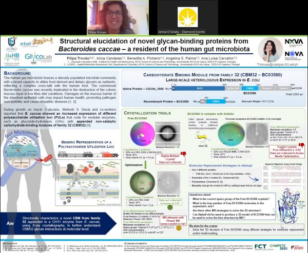
Best Poster Award at Diamond-CCP4 Workshop to Filipa Trovão!

Congratulations to our PhD student Filipa Trovão for winning the Best Poster Award at the Diamond-CCP4 Data Collection and Structure Solution Workshop (https://www.ccp4.ac.uk/schools/DLS-2021)!

The workshop covered all aspects of 3D structure solution, from data collection through to phasing, refinement and validation, lectured by exp

Public presentation of the PhD Thesis Plan of Filipa Trovão

Today, Filipa Trovão, a joint PhD student of Angelina Palma (GlycoLab), Ana Luísa Carvalho (XTAL) and Yan Liu (Imperial College London), has publicly presented the Thesis Plan of her PhD work.

Well done, Filipa! Your presentation was very clear and the Thesis Advisory Committee members (Paula Videira and Pedro Bule) acknowledged that your work is on the right track!

In the European Researchers' Night 2021

As usual, we are part of this great celebration of all those that work and love science!

https://noitedosinvestigadores.org/programa-online/

This year, the event is dedicated to Climate and 3 of our projects meet this subject perfectly.

Goodfellow, BJ, Freire F, Carvalho AL, Aveiro SS, Charbonnier P, Moulis J-M, Delgado L, Ferreira GC, Rodrigues JE, Poussin-Courmontagne P, Birck C, McEwen A, Macedo AL.  2021.  The SOUL family of heme-binding proteins: Structure and function 15 years later, 2021. 448:214189. AbstractWebsite

The SOUL, or heme-binding protein HBP/SOUL, family represents a group of evolutionary conserved putative heme-binding proteins that contains a number of members in animal, plant andbacterial species. The structures of the murine form of HEBP1, or p22HBP, and the human form of HEBP2, or SOUL, have been determined in 2006 and 2011 respectively. In this work we discuss the structures of HEBP1 and HEBP2 in light of new X-ray data for heme bound murine HEBP1. The interaction between tetrapyrroles and HEBP1, initially proven to be hydrophobic in nature, was thought to also involve electrostatic interactions between heme propionate groups and positively charged amino acid side chains. However, the new X-ray structure, and results from murine HEBP1 variants and human HEBP1, confirm the hydrophobic nature of the heme-HEBP1 interaction, resulting in Kd values in the low nanomolar range, and rules out any electrostatic stabilization. Results from NMR relaxation time measurements for human HEBP1 describe a rigid globular protein with no change in motional regime upon heme binding. X-ray structures deposited in the PDB for human HEBP2 are very similar to each other and to the new heme-bound murine HEBP1 X-ray structure (backbone rmsd ca. 1 Å). Results from a HSQC spectrum centred on the histidine side chain Nδ-proton region for HEBP2 confirm that HEBP2 does not bind heme via H42 as no chemical shift differences were observed upon heme addition for backbone NH and Nδ protons. A survey of the functions attributed to HEBP1 and HEBP2 over the last 20 years span a wide range of cellular pathways. Interestingly, many of them are specific to higher eukaryotes, particularly mammals and a potential link between heme release under oxidative stress and human HEBP1 is also examined using recent data. However, at the present moment, trying to relate function to the involvement of heme or tetrapyrrole binding, specifically, makes little sense with our current biological knowledge and can only be applied to HEBP1, as HEBP2 does not interact with heme. We suggest that it may not be justified to call this very small family of proteins, heme-binding proteins. The family may be more correctly called “the SOUL family of proteins related to cellular fate” as, even though only HEBP1 binds heme tightly, both proteins may be involved in cell survival and/or proliferation.

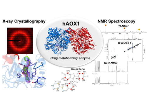
Mota, C, Diniz A, Coelho C, Santos-Silva T, Esmaeeli M, Leimkühler S, Cabrita EJ, Marcelo F, Romão MJ.  2021.  Interrogating the Inhibition Mechanisms of Human Aldehyde Oxidase by X-ray Crystallography and NMR Spectroscopy: The Raloxifene Case, 2021. Journal of Medicinal ChemistryJournal of Medicinal Chemistry. : American Chemical Society AbstractWebsite

Human aldehyde oxidase (hAOX1) is mainly present in the liver and has an emerging role in drug metabolism, since it accepts a wide range of molecules as substrates and inhibitors. Herein, we employed an integrative approach by combining NMR, X-ray crystallography, and enzyme inhibition kinetics to understand the inhibition modes of three hAOX1 inhibitors—thioridazine, benzamidine, and raloxifene. These integrative data indicate that thioridazine is a noncompetitive inhibitor, while benzamidine presents a mixed type of inhibition. Additionally, we describe the first crystal structure of hAOX1 in complex with raloxifene. Raloxifene binds tightly at the entrance of the substrate tunnel, stabilizing the flexible entrance gates and elucidating an unusual substrate-dependent mechanism of inhibition with potential impact on drug–drug interactions. This study can be considered as a proof-of-concept for an efficient experimental screening of prospective substrates and inhibitors of hAOX1 relevant in drug discovery.Human aldehyde oxidase (hAOX1) is mainly present in the liver and has an emerging role in drug metabolism, since it accepts a wide range of molecules as substrates and inhibitors. Herein, we employed an integrative approach by combining NMR, X-ray crystallography, and enzyme inhibition kinetics to understand the inhibition modes of three hAOX1 inhibitors—thioridazine, benzamidine, and raloxifene. These integrative data indicate that thioridazine is a noncompetitive inhibitor, while benzamidine presents a mixed type of inhibition. Additionally, we describe the first crystal structure of hAOX1 in complex with raloxifene. Raloxifene binds tightly at the entrance of the substrate tunnel, stabilizing the flexible entrance gates and elucidating an unusual substrate-dependent mechanism of inhibition with potential impact on drug–drug interactions. This study can be considered as a proof-of-concept for an efficient experimental screening of prospective substrates and inhibitors of hAOX1 relevant in drug discovery.

dos Santos, R, Romão MJ, Roque ACA, Carvalho AL.  2021.  Magnetic particles used in a new approach for designed protein crystallization. CrystEngComm. 23:1083-1090.: The Royal Society of Chemistry AbstractWebsite

After more than one hundred and thirty thousand protein structures determined by X-ray crystallography{,} the challenge of protein crystallization for 3D structure determination remains. In the quest for additives for efficient protein crystallization{,} inorganic materials emerge as an alternative. Magnetic particles (MPs) are versatile inorganic materials{,} easy to use{,} modify and manipulate in a wide range of biological assays. The potential of using functionalised MPs as crystallization chaperones for protein crystallization was shown in this work. MPs with distinct coatings were rationally designed to promote protein crystallization by affinity-triggered heterogeneous nucleation. Hen egg white lysozyme (HEWL) and trypsin{,} were crystallized in the presence of MPs either bare or coated with a polysaccharide (chitin) or a protein (casein){,} respectively. The addition of MPs was characterized in terms of bound protein to the MPs{,} crystal morphology{,} time-lapse of crystal emergence{,} crystallization yield fold change and crystal diffraction quality for structure determination. The MPs additives have shown to bind to the respective target protein{,} and to promote nucleation and crystal growth without compromising crystal morphology. On the other hand{,} MPs addition led to faster detectable crystal emergence and up to 13 times higher crystallization yield{,} addressing some the challenges in protein crystallization{,} the main bottleneck of macromolecular crystallography. Structure determination of the protein crystallized in the presence of MPs revealed that the structural characteristics of the protein remained unchanged{,} as shown by the superposition with PDB annotated proteins. Moreover{,} and unlike most reported cases{,} it was possible to exclude the inhibitor benzamidine during trypsin crystallisation{,} which is a remarkable result opening new prospects in enzyme engineering and drug design. Our results show that MPs coated with affinity ligands to target proteins can be used as controlled and tailor-made crystallization inducers.

Polino, M, Rho HS, Pina MP, Mallada R, Carvalho AL, Romão MJ, Coelhoso I, Gardeniers JGE, Crespo JG, Portugal CAM.  2021.  Protein Crystallization in a Microfluidic Contactor with Nafion®117 Membranes. Membranes. 11, Number 8 AbstractWebsite

Protein crystallization still remains mostly an empirical science, as the production of crystals with the required quality for X-ray analysis is dependent on the intensive screening of the best protein crystallization and crystal’s derivatization conditions. Herein, this demanding step was addressed by the development of a high-throughput and low-budget microfluidic platform consisting of an ion exchange membrane (117 Nafion® membrane) sandwiched between a channel layer (stripping phase compartment) and a wells layer (feed phase compartment) forming 75 independent micro-contactors. This microfluidic device allows for a simultaneous and independent screening of multiple protein crystallization and crystal derivatization conditions, using Hen Egg White Lysozyme (HEWL) as the model protein and Hg2+ as the derivatizing agent. This microdevice offers well-regulated crystallization and subsequent crystal derivatization processes based on the controlled transport of water and ions provided by the 117 Nafion® membrane. Diffusion coefficients of water and the derivatizing agent (Hg2+) were evaluated, showing the positive influence of the protein drop volume on the number of crystals and crystal size. This microfluidic system allowed for crystals with good structural stability and high X-ray diffraction quality and, thus, it is regarded as an efficient tool that may contribute to the enhancement of the proteins’ crystals structural resolution.

Ferreira, P, Cerqueira NSMFA, Fernandes PA, Romão MJ, Ramos MJ.  2020.  Catalytic Mechanism of Human Aldehyde Oxidase, 2020. ACS CatalysisACS Catalysis. 10(16):9276-9286.: American Chemical Society AbstractWebsite

The mechanism of oxidation of N-heterocycle phthalazine to phthalazin-1(2H)-one and its associated free energy profile, catalyzed by human aldehyde oxidase (hAOX1), was studied in atomistic detail using QM/MM methodologies. The studied reaction was found to involve three sequential steps: (i) protonation of the substrate’s N2 atom by Lys893, (ii) nucleophilic attack of the hydroxyl group of the molybdenum cofactor (Moco) to the substrate, and (iii) hydride transfer from the substrate to the sulfur atom of the Moco. The free energy profile that was calculated revealed that the rate-limiting step corresponds to hydride transfer. It was also found that Lys893 plays a relevant role in the reaction, being important not only for the anchorage of the substrate close to the Moco, but also in the catalytic reaction. The variations of the oxidation state of the molybdenum ion throughout the catalytic cycle were examined too. We found out that during the displacement of the products away from the Moco, the transfer of electrons from the catalytic site to the FAD site was proton-coupled. As a consequence, the most favorable and fastest pathway for the enzyme to complete its catalytic cycle was that with MoV and a deprotonated SH ligand of the Moco with the FAD molecule converted to its semiquinone form, FADH•.The mechanism of oxidation of N-heterocycle phthalazine to phthalazin-1(2H)-one and its associated free energy profile, catalyzed by human aldehyde oxidase (hAOX1), was studied in atomistic detail using QM/MM methodologies. The studied reaction was found to involve three sequential steps: (i) protonation of the substrate’s N2 atom by Lys893, (ii) nucleophilic attack of the hydroxyl group of the molybdenum cofactor (Moco) to the substrate, and (iii) hydride transfer from the substrate to the sulfur atom of the Moco. The free energy profile that was calculated revealed that the rate-limiting step corresponds to hydride transfer. It was also found that Lys893 plays a relevant role in the reaction, being important not only for the anchorage of the substrate close to the Moco, but also in the catalytic reaction. The variations of the oxidation state of the molybdenum ion throughout the catalytic cycle were examined too. We found out that during the displacement of the products away from the Moco, the transfer of electrons from the catalytic site to the FAD site was proton-coupled. As a consequence, the most favorable and fastest pathway for the enzyme to complete its catalytic cycle was that with MoV and a deprotonated SH ligand of the Moco with the FAD molecule converted to its semiquinone form, FADH•.