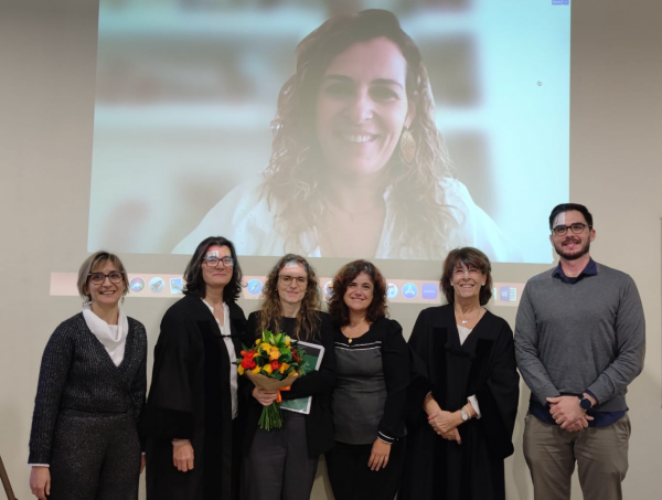
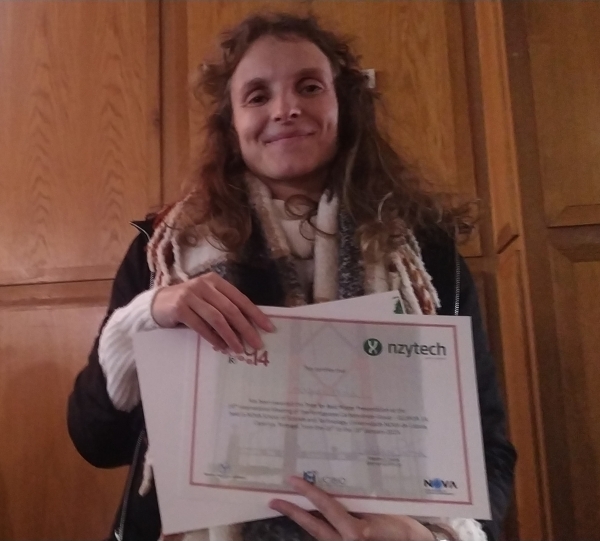
xtal

Dias, AMGC, Moreira IP, Lychko I, Lopes Soares C, Nurrito A, Moura Barbosa AJ, Lutz-Bueno V, Mezzenga R, Carvalho AL, Pina AS, Roque ACA.  2023.  Hierarchical self-assembly of a reflectin-derived peptide. Frontiers in Chemistry. 11 AbstractWebsite

Reflectins are a family of intrinsically disordered proteins involved in cephalopod camouflage, making them an interesting source for bioinspired optical materials. Understanding reflectin assembly into higher-order structures by standard biophysical methods enables the rational design of new materials, but it is difficult due to their low solubility. To address this challenge, we aim to understand the molecular self-assembly mechanism of reflectin’s basic unit—the protopeptide sequence YMDMSGYQ—as a means to understand reflectin’s assembly phenomena. Protopeptide self-assembly was triggered by different environmental cues, yielding supramolecular hydrogels, and characterized by experimental and theoretical methods. Protopeptide films were also prepared to assess optical properties. Our results support the hypothesis for the protopeptide aggregation model at an atomistic level, led by hydrophilic and hydrophobic interactions mediated by tyrosine residues. Protopeptide-derived films were optically active, presenting diffuse reflectance in the visible region of the light spectrum. Hence, these results contribute to a better understanding of the protopeptide structural assembly, crucial for the design of peptide- and reflectin-based functional materials.

Silva, JM, Cerofolini L, Carvalho AL, Ravera E, Fragai M, Parigi G, Macedo AL, Geraldes CFGC, Luchinat C.  2023.  Elucidating the concentration-dependent effects of thiocyanate binding to carbonic anhydrase, 2023. 244:112222. AbstractWebsite

Many proteins naturally carry metal centers, with a large share of them being in the active sites of several enzymes. Paramagnetic effects are a powerful source of structural information and, therefore, if the native metal is paramagnetic, or it can be functionally substituted with a paramagnetic one, paramagnetic effects can be used to study the metal sites, as well as the overall structure of the protein. One notable example is cobalt(II) substitution for zinc(II) in carbonic anhydrase. In this manuscript we investigate the effects of sodium thiocyanate on the chemical environment of the metal ion of the human carbonic anhydrase II. The electron paramagnetic resonance (EPR) titration of the cobalt(II) protein with thiocyanate shows that the EPR spectrum changes from A-type to C-type on passing from 1:1 to 1:1000-fold ligand excess. This indicates the occurrence of a change in the electronic structure, which may reflect a sizable change in the metal coordination environment in turn caused by a modification of the frozen solvent glass. However, paramagnetic nuclear magnetic resonance (NMR) data indicate that the metal coordination cage remains unperturbed even in 1:1000-fold ligand excess. This result proves that the C-type EPR spectrum observed at large ligand concentration should be ascribed to the low temperature at which EPR measurements are performed, which impacts on the structure of the protein when it is destabilized by a high concentration of a chaotropic agent.

Nóbrega, CS, Carvalho AL, Romão MJ, Pauleta SR.  2023.  Structural Characterization of Neisseria gonorrhoeae Bacterial Peroxidase—Insights into the Catalytic Cycle of Bacterial Peroxidases. International Journal of Molecular Sciences. 24, Number 7 AbstractWebsite

Neisseria gonorrhoeae is an obligate human pathogenic bacterium responsible for gonorrhea, a sexually transmitted disease. The bacterial peroxidase, an enzyme present in the periplasm of this bacterium, detoxifies the cells against hydrogen peroxide and constitutes one of the primary defenses against exogenous and endogenous oxidative stress in this organism. The 38 kDa heterologously produced bacterial peroxidase was crystallized in the mixed-valence state, the active state, at pH 6.0, and the crystals were soaked with azide, producing the first azide-inhibited structure of this family of enzymes. The enzyme binds exogenous ligands such as cyanide and azide, which also inhibit the catalytic activity by coordinating the P heme iron, the active site, and competing with its substrate, hydrogen peroxide. The inhibition constants were estimated to be 0.4 ± 0.1 µM and 41 ± 5 mM for cyanide and azide, respectively. Imidazole also binds and inhibits the enzyme in a more complex mechanism by binding to P and E hemes, which changes the reduction potential of the latest heme. Based on the structures now reported, the catalytic cycle of bacterial peroxidases is revisited. The inhibition studies and the crystal structure of the inhibited enzyme comprise the first platform to search and develop inhibitors that target this enzyme as a possible new strategy against N. gonorrhoeae.

XTAL Twitter

dvefverv 

Correia, VG, Trovão F, Pinheiro BA, Brás JLA, Silva LM, Nunes C, Coimbra MA, Liu Y, Feizi T, Fontes CMGA, Mulloy B, Chai W, Carvalho AL, Palma AS.  2021.  Mapping Molecular Recognition of β1,3-1,4-Glucans by a Surface Glycan-Binding Protein from the Human Gut Symbiont Bacteroides ovatus, December. Microbiology spectrum. 9:e0182621., Number 3 AbstractWebsite

A multigene polysaccharide utilization locus (PUL) encoding enzymes and surface carbohydrate (glycan)-binding proteins (SGBPs) was recently identified in prominent members of <i>Bacteroidetes</i> in the human gut and characterized in Bacteroides ovatus. This PUL-encoded system specifically targets mixed-linkage β1,3-1,4-glucans, a group of diet-derived carbohydrates that promote a healthy microbiota and have potential as prebiotics. The BoSGBP<sub>MLG</sub>-A protein encoded by the <i>BACOVA_2743</i> gene is a SusD-like protein that plays a key role in the PUL's specificity and functionality. Here, we perform a detailed analysis of the molecular determinants underlying carbohydrate binding by BoSGBP<sub>MLG</sub>-A, combining carbohydrate microarray technology with quantitative affinity studies and a high-resolution X-ray crystallography structure of the complex of BoSGBP<sub>MLG</sub>-A with a β1,3-1,4-nonasaccharide. We demonstrate its unique binding specificity toward β1,3-1,4-gluco-oligosaccharides, with increasing binding affinities up to the octasaccharide and dependency on the number and position of β1,3 linkages. The interaction is defined by a 41-Å-long extended binding site that accommodates the oligosaccharide in a mode distinct from that of previously described bacterial β1,3-1,4-glucan-binding proteins. In addition to the shape complementarity mediated by CH-π interactions, a complex hydrogen bonding network complemented by a high number of key ordered water molecules establishes additional specific interactions with the oligosaccharide. These support the twisted conformation of the β-glucan backbone imposed by the β1,3 linkages and explain the dependency on the oligosaccharide chain length. We propose that the specificity of the PUL conferred by BoSGBP<sub>MLG</sub>-A to import long β1,3-1,4-glucan oligosaccharides to the bacterial periplasm allows <i>Bacteroidetes</i> to outcompete bacteria that lack this PUL for utilization of β1,3-1,4-glucans. <b>IMPORTANCE</b> With the knowledge of bacterial gene systems encoding proteins that target dietary carbohydrates as a source of nutrients and their importance for human health, major efforts are being made to understand carbohydrate recognition by various commensal bacteria. Here, we describe an integrative strategy that combines carbohydrate microarray technology with structural studies to further elucidate the molecular determinants of carbohydrate recognition by BoSGBP<sub>MLG</sub>-A, a key protein expressed at the surface of Bacteroides ovatus for utilization of mixed-linkage β1,3-1,4-glucans. We have mapped at high resolution interactions that occur at the binding site of BoSGBP<sub>MLG</sub>-A and provide evidence for the role of key water-mediated interactions for fine specificity and affinity. Understanding at the molecular level how commensal bacteria, such as prominent members of <i>Bacteroidetes</i>, can differentially utilize dietary carbohydrates with potential prebiotic activities will shed light on possible ways to modulate the microbiome to promote human health.

Esteves, C, Palma SICJ, Costa HMA, Alves C, Santos GMC, Ramou E, Carvalho AL, Alves V, Roque ACA.  2022.  Tackling Humidity with Designer Ionic Liquid-Based Gas Sensing Soft Materials. Advanced Materials. 34:2107205., Number 8 AbstractWebsite

Abstract Relative humidity is simultaneously a sensing target and a contaminant in gas and volatile organic compound (VOC) sensing systems, where strategies to control humidity interference are required. An unmet challenge is the creation of gas-sensitive materials where the response to humidity is controlled by the material itself. Here, humidity effects are controlled through the design of gelatin formulations in ionic liquids without and with liquid crystals as electrical and optical sensors, respectively. In this design, the anions [DCA]− and [Cl]− of room temperature ionic liquids from the 1-butyl-3-methylimidazolium family tailor the response to humidity and, subsequently, sensing of VOCs in dry and humid conditions. Due to the combined effect of the materials formulations and sensing mechanisms, changing the anion from [DCA]− to the much more hygroscopic [Cl]−, leads to stronger electrical responses and much weaker optical responses to humidity. Thus, either humidity sensors or humidity-tolerant VOC sensors that do not require sample preconditioning or signal processing to correct humidity impact are obtained. With the wide spread of 3D- and 4D-printing and intelligent devices, the monitoring and tuning of humidity in sustainable biobased materials offers excellent opportunities in e-nose sensing arrays and wearable devices compatible with operation at room conditions.

Duarte, M, Alves VD, Correia M, Caseiro C, Ferreira LMA, Romão MJ, Carvalho AL, Najmudin S, Bayer EA, Fontes CMGA, Bule P.  2023.  Structure-function studies can improve binding affinity of cohesin-dockerin interactions for multi-protein assemblies, 2023. 224:55-67. AbstractWebsite

The cellulosome is an elaborate multi-enzyme structure secreted by many anaerobic microorganisms for the efficient degradation of lignocellulosic substrates. It is composed of multiple catalytic and non-catalytic components that are assembled through high-affinity protein-protein interactions between the enzyme-borne dockerin (Doc) modules and the repeated cohesin (Coh) modules present in primary scaffoldins. In some cellulosomes, primary scaffoldins can interact with adaptor and cell-anchoring scaffoldins to create structures of increasing complexity. The cellulosomal system of the ruminal bacterium, Ruminococcus flavefaciens, is one of the most intricate described to date. An unprecedent number of different Doc specificities results in an elaborate architecture, assembled exclusively through single-binding-mode type-III Coh-Doc interactions. However, a set of type-III Docs exhibits certain features associated with the classic dual-binding mode Coh-Doc interaction. Here, the structure of the adaptor scaffoldin-borne ScaH Doc in complex with the Coh from anchoring scaffoldin ScaE is described. This complex, unlike previously described type-III interactions in R. flavefaciens, was found to interact in a dual-binding mode. The key residues determining Coh recognition were also identified. This information was used to perform structure-informed protein engineering to change the electrostatic profile of the binding surface and to improve the affinity between the two modules. The results show that the nature of the residues in the ligand-binding surface plays a major role in Coh recognition and that Coh-Doc affinity can be manipulated through rational design, a key feature for the creation of designer cellulosomes or other affinity-based technologies using tailored Coh-Doc interactions.

Santos, MFA, Sciortino G, Correia I, Fernandes ACP, Santos-Silva T, Pisanu F, Garribba E, Pessoa JC.  2022.  Binding of VIVO2+, VIVOL, VIVOL2 and VVO2L Moieties to Proteins: X-ray/Theoretical Characterization and Biological Implications, 2022. Chemistry – A European JournalChemistry – A European Journal. 28(40):e202200105.: John Wiley & Sons, Ltd AbstractWebsite

Abstract Vanadium compounds have frequently been proposed as therapeutics, but their application has been hampered by the lack of information on the different V-containing species that may form and how these interact with blood and cell proteins, and with enzymes. Herein, we report several resolved crystal structures of lysozyme with bound VIVO2+ and VIVOL2+, where L=2,2?-bipyridine or 1,10-phenanthroline (phen), and of trypsin with VIVO(picolinato)2 and VVO2(phen)+ moieties. Computational studies complete the refinement and shed light on the relevant role of hydrophobic interactions, hydrogen bonds, and microsolvation in stabilizating the structure. Noteworthy is that the trypsin?VVO2(phen) and trypsin?VIVO(OH)(phen) adducts correspond to similar energies, thus suggesting a possible interconversion under physiological/biological conditions. The obtained data support the relevance of hydrolysis of VIV and VV complexes in the several types of binding established with proteins and the formation of different adducts that might contribute to their pharmacological action, and significantly widen our knowledge of vanadium?protein interactions.

Trovão, F, Correia VG, Lourenço FM, Ribeiro DO, Carvalho AL, Palma AS, Pinheiro BA.  2023.  The structure of a Bacteroides thetaiotamicron carbohydrate-binding module provides new insight into the recognition of complex pectic polysaccharides by the human microbiome, 2023. :100084. AbstractWebsite

TheBacteroides thetaiotaomicronhas developed a consortium of enzymes capable of overcoming steric constraints and degrading, in a sequential manner, the complex rhamnogalacturonan II (RG-II) polysaccharide. BT0996 protein acts in the initial stages of the RGII depolymerisation, where its two catalytic modules remove the terminal monosaccharides from RG-II side chains A and B. BT0996 is modular and has three putative carbohydrate-binding modules (CBMs) for which the roles in the RG-II degradation are unknown. Here, we present the characterisation of themoduleat the C-terminal domain, which we designated BT0996C. The high-resolution structure obtained by X-ray crystallography reveals that the protein displays a typical β-sandwich fold with structural similarity to CBMs assigned to families 6 and 35. The distinctive features are: 1) the presence of several charged residues at the BT0996-C surface creating a large, broad positive lysine-rich patch that encompasses the putative binding site; and 2) the absence of the highly conserved binding-site signatures observed in CBMs from families 6 and 35, such as region A tryptophan and region C asparagine. These findings hint at a binding mode of BT0996-C not yet observed in its homologues. In line with this, carbohydrate microarrays and microscale thermophoresis show the ability of BT0996-C to bind α1-4-linked polygalacturonic acid, and that electrostatic interactions are essential for the recognition of the anionic polysaccharide. The results support the hypothesis that BT0996-C may have evolved to potentiate the action of BT0996 catalytic modules on the complex structure of RG-II by binding to the polygalacturonic acid backbone sequence.

Congratulations to Alicia Candeias!

Congratulations to our student Alicia Candeias for successfully defending her MSc thesis in Biochemistry at NOVA School of Science and Technology.
Her thesis entitled "A molecular view on how a commensal bacterium thrives in the human gut" was supervised by Ana Luísa Carvalho, from XTAL, and Angelina Sá Palma, from the GlycoLab.

Congratulations, Doctor Francisco!

Congratulations to Francisco Leisico for successfully concluding his PhD Thesis, today!
His thesis is entitled "Unravelling the reaction mechanism of glutamate amidation in Staphylococcus aureus peptidoglycan", supervised by Teresa Santos-Silva and Maria João Romão.
Our best wishes for your future, Francisco!
Here's a photo of the nice, in person, lunch party!
Moreira, IP, Esteves C, Palma SICJ, Ramou E, Carvalho ALM, Roque ACA.  2022.  Synergy between silk fibroin and ionic liquids for active gas-sensing materials. Materials Today Bio. :100290. AbstractWebsite

Silk fibroin is a biobased material with excellent biocompatibility and mechanical properties, but its use in bioelectronics is hampered by the difficult dissolution and low intrinsic conductivity. Some ionic liquids are known to dissolve fibroin but removed after fibroin processing. However, ionic liquids and fibroin can cooperatively give rise to functional materials, and there are untapped opportunities in this combination. The dissolution of fibroin, followed by gelation, in designer ionic liquids from the imidazolium chloride family with varied alkyl chain lengths (2–10 carbons) is shown here. The alkyl chain length of the anion has a large impact on fibroin secondary structure which adopts unconventional arrangements, yielding robust gels with distinct hierarchical organization. Furthermore, and due to their remarkable air-stability and ionic conductivity, fibroin ionogels are exploited as active electrical gas sensors in an electronic nose revealing the unravelled possibilities of fibroin in soft and flexible electronics.

Barroca-Ferreira, J, Cruz-Vicente P, Santos MFA, Rocha SM, Santos-Silva T, Maia CJ, Passarinha LA.  2021.  Enhanced Stability of Detergent-Free Human Native STEAP1 Protein from Neoplastic Prostate Cancer Cells upon an Innovative Isolation Procedure. International Journal of Molecular Sciences. 22, Number 18 AbstractWebsite

Background: The STEAP1 is a cell-surface antigen over-expressed in prostate cancer, which contributes to tumor progression and aggressiveness. However, the molecular mechanisms underlying STEAP1 and its structural determinants remain elusive. Methods: The fraction capacity of Butyl- and Octyl-Sepharose matrices on LNCaP lysates was evaluated by manipulating the ionic strength of binding and elution phases, followed by a Co-Immunoprecipitation (Co-IP) polishing. Several potential stabilizing additives were assessed, and the melting temperature (Tm) values ranked the best/worst compounds. The secondary structure of STEAP1 was identified by circular dichroism. Results: The STEAP1 was not fully captured with 1.375 M (Butyl), in contrast with interfering heterologous proteins, which were strongly retained and mostly eluted with water. This single step demonstrated higher selectivity of Butyl-Sepharose for host impurities removal from injected crude samples. Co-IP allowed recovering a purified fraction of STEAP1 and contributed to unveil potential physiologically interacting counterparts with the target. A Tm of  55 °C was determined, confirming STEAP1 stability in the purification buffer. A predominant α-helical structure was identified, ensuring the protein’s structural stability. Conclusions: A method for successfully isolating human STEAP1 from LNCaP cells was provided, avoiding the use of detergents to achieve stability, even outside a membrane-mimicking environment.