Confocal Microscopy
Confocal microscopy is a fluorescence optical imaging technique for increasing optical resolution and contrast of a micrograph. This technique uses a laser point source to scan the sample and a pinhole to reduce collection of light from outside the focal plane and allows investigators to acquire thin optical sections at various focal planes. Stacks of images can be acquired for 3D visualization.
Application examples:
- Life Sciences: wide range of imaging applications including the study of the spatial distributions of macromolecules in either fixed or living cells (e.g., protein localization analysis), automated collection of 3D data, the imaging of multiple labeled specimens and the measurements of physiological events in living cells (e.g., cell cycle studies).
- Material Sciences: used for investigating polymers, coatings, emulsions, etc.
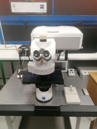
We operate with the Zeiss LSM 710, a confocal laser scanning microscope able to generate high-resolution 3D images of specimens with high sensitivity and low photodamage.
System components:
LASERs
|
Laser Unit |
Wavelength |
Maximum Power |
|
Diode 405-30 |
405 nm |
30 mW |
|
Argon |
458, 488 and 514 nm |
25 mW |
|
DPSS 561-10 |
561 nm |
15 mW |
|
HeNe633 |
633 nm |
5 mW |
Objectives
|
Magnification |
Model |
Immersion |
|
10x |
EC Plan-Neofluar |
air |
|
40x |
EC Plan-Neofluar |
oil |
|
63x |
Plan-Apochromat |
oil |
Filter sets (ocular)
|
Reflectors |
Position/name |
Excitation |
Dichroic |
Emission |
|
Green |
1-38 GFP |
450-490 nm |
495 nm |
500-550 nm |
|
Red |
2-43 DsRed |
533-558 nm |
570 nm |
570-640 nm |
|
Blue |
3-49 DAPI |
G 365 nm |
395 nm |
420-470 nm |
Software
Image acquisition and analysis is performed using the Zen 2010 software that includes the following modules:
- Z stack
- Time Series
- Bleaching
- Tile Scan
- Positions
- Regions
Publications with our Zeiss LSM 710:
Araújo, D. et al. Chitin-Glucan Complex Hydrogels: Physical-Chemical Characterization, Stability, In Vitro Drug Permeation, and Biological Assessment in Primary Cells. Polymers (Basel). 15, (2023).
https://doi.org/10.3390/polym15040791
Confocal Microscopy
Confocal microscopy is a fluorescence optical imaging technique for increasing optical resolution and contrast of a micrograph. This technique uses a laser point source to scan the sample and a pinhole to reduce collection of light from outside the focal plane and allows investigators to acquire thin optical sections at various focal planes. Stacks of images can be acquired for 3D visualization.
Application examples:
- Life Sciences: wide range of imaging applications including the study of the spatial distributions of macromolecules in either fixed or living cells (e.g., protein localization analysis), automated collection of 3D data, the imaging of multiple labeled specimens and the measurements of physiological events in living cells (e.g., cell cycle studies).
- Material Sciences: used for investigating polymers, coatings, emulsions, etc.
We operate with the Zeiss LSM 710, a confocal laser scanning microscope able to generate high-resolution 3D images of specimens with high sensitivity and low photodamage.
System components:
LASERs
|
Laser Unit |
Wavelength |
Maximum Power |
|
Diode 405-30 |
405 nm |
30 mW |
|
Argon |
458, 488 and 514 nm |
25 mW |
|
DPSS 561-10 |
561 nm |
15 mW |
|
HeNe633 |
633 nm |
5 mW |
Objectives
|
Magnification |
Model |
Immersion |
|
10x |
EC Plan-Neofluar |
air |
|
40x |
EC Plan-Neofluar |
oil |
|
63x |
Plan-Apochromat |
oil |
Filter sets (ocular)
|
Reflectors |
Position/name |
Excitation |
Dichroic |
Emission |
|
Green |
1-38 GFP |
450-490 nm |
495 nm |
500-550 nm |
|
Red |
2-43 DsRed |
533-558 nm |
570 nm |
570-640 nm |
|
Blue |
3-49 DAPI |
G 365 nm |
395 nm |
420-470 nm |
Software
Image acquisition and analysis is performed using the Zen 2010 software that includes the following modules:
- Z stack
- Time Series
- Bleaching
- Tile Scan
- Positions
- Regions
Publications with our Zeiss LSM 710:
Araújo, D. et al. Chitin-Glucan Complex Hydrogels: Physical-Chemical Characterization, Stability, In Vitro Drug Permeation, and Biological Assessment in Primary Cells. Polymers (Basel). 15, (2023).
https://doi.org/10.3390/polym15040791